Raman Imaging for Advanced Scientific and Industrial Applications
Raman Imaging for Advanced Scientific and Industrial Applications
Raman Imaging has become a powerful and trusted technique in modern analytical science. It offers a non-destructive and label-free way to identify chemical composition and visualize molecular structures at microscopic and even nanoscopic scales. By combining Raman spectroscopy with advanced imaging systems, researchers can study molecular makeup, subtle structural variations, and complex chemical interactions within a wide range of samples.
Today, Raman Imaging plays an important role across multiple fields, including materials science, pharmaceuticals, biomedical research, semiconductor engineering, polymer science, geology, and nanotechnology. Its ability to generate detailed chemical maps without altering the sample makes it especially valuable in both research and industrial environments.
Unlike conventional imaging techniques that rely on indirect indicators, Raman Imaging directly links every point in the image to a unique molecular vibrational signature. This direct chemical insight ensures high accuracy while maintaining sample integrity—no dyes, no damage, and no compromise.
Fundamental Principles of Raman Imaging
Raman Imaging is based on the Raman effect, which occurs when monochromatic laser light interacts with a material and undergoes inelastic scattering. During this interaction, a small portion of the scattered light experiences a shift in energy that corresponds to the vibrational and structural characteristics of the molecules present.
These energy shifts act as molecular fingerprints. When collected across thousands or even millions of spatial points, they form a hyperspectral dataset where each pixel contains complete spectral information. This allows researchers to visualize chemical distributions and structural features with exceptional precision.
Key components of the process include controlled laser excitation, accurate detection of vibrational spectra, spatial mapping, and advanced data interpretation techniques.
Raman Imaging Techniques and Modalities
Confocal Raman Imaging
Confocal Raman microscopy is widely used when high spatial resolution and depth profiling are required. By eliminating out-of-focus signals, it provides clear three-dimensional insights into thin films, layered structures, and biological samples.
Widefield Raman Imaging
For applications that demand faster data acquisition over larger areas, widefield Raman Imaging is an effective solution. It is commonly used in industrial inspection and quality control, where rapid surface analysis is essential.
TERS- Tip-Enhanced Raman Scattering
Tip-Enhanced Raman Imaging extends Raman analysis into the nanoscale domain, achieving spatial resolution below 10 nanometers. By integrating Raman spectroscopy with scanning probe techniques, TERS enables detailed investigation of nanomaterials, plasmonic structures, and semiconductor devices.
Coherent Raman Imaging
Techniques such as Coherent Anti-Stokes Raman Scattering (CARS) and Stimulated Raman Scattering (SRS) enhance signal strength and allow real-time imaging. These methods are particularly useful in live-cell studies and dynamic biomedical research.
Key Advantages of Raman Imaging
Raman Imaging offers a unique combination of benefits that few analytical techniques can match. It is completely label-free and non-destructive, requires minimal sample preparation, and provides highly specific chemical information. The technique works effectively with solids, liquids, and gases, delivering high spatial and spectral resolution across diverse sample types.
Because it can capture both structural and compositional information simultaneously, Raman Imaging is equally valuable in exploratory research and industrial quality assurance.
Applications Across Industries
Biomedical and Life Sciences - In biomedical research, Raman Imaging enables the differentiation between healthy and diseased tissues, detection of biochemical changes at the cellular level, and detailed monitoring of drug–cell interactions.
Pharmaceutical Industry - Raman Imaging plays a crucial role in pharmaceutical quality control. It helps verify the uniform distribution of active pharmaceutical ingredients, identify different polymorphic forms, and detect trace-level impurities.
Materials Science and Nanotechnology - From polymers and ceramics to graphene and advanced composites, Raman Imaging provides insights into crystallinity, stress distribution, phase transitions, and material defects.
Semiconductors and Electronics - In semiconductor manufacturing, Raman Imaging is used to visualize strain, assess layer thickness, and locate defects. These capabilities support wafer inspection, device reliability testing, and advanced packaging technologies.
Geology and Mineralogy - Raman Imaging allows non-invasive mineral identification and geochemical mapping. Its ability to analyze rare or valuable specimens without physical alteration makes it especially valuable in geological research.
Request for Information
Do you have any questions or requests? Use this form to contact our team.
Data Analysis and Interpretation
The true strength of Raman Imaging lies not only in data collection but also in data interpretation. Advanced analytical tools such as Principal Component Analysis (PCA), Cluster Analysis, Multivariate Curve Resolution, and machine learning techniques are used to extract meaningful insights from complex spectral datasets. These methods help resolve overlapping signals, identify hidden patterns, and enable accurate quantification.
IndiRAM Raman Imaging
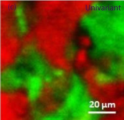
a.) Raman Imaging of Pharmaceutical API's distribution in a Tablet -
Distribution of Ibuprofen API in Red and Paracetamol APIs in Green | b.) Material Science - 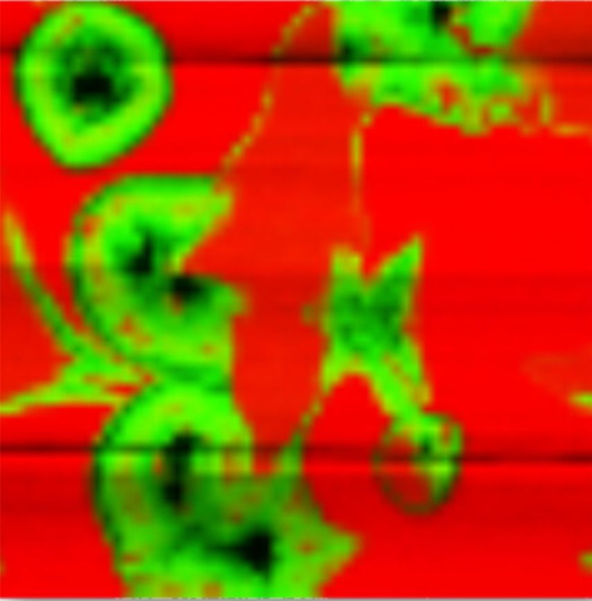 Red color is Silicon and Green is MoS2 |
Instrumentation and System Design
A high-performance Raman Imaging system integrates stable laser sources (ranging from UV to near-infrared), high-resolution spectrometers, and sensitive CCD detectors. Precision motorized stages and advanced software platforms support accurate imaging and efficient data analysis. System configurations are carefully optimized to meet specific application requirements related to resolution, sensitivity, and acquisition speed.
Technical Challenges and Optimization Strategies
Despite its versatility, Raman Imaging presents certain technical challenges, including weak signal intensity, fluorescence interference, and longer acquisition times. These issues are addressed through optimized wavelength selection, advanced optical filtering, improved detector sensitivity, computational noise reduction, and automated mapping workflows. Such strategies ensure reliable and reproducible results.
Future Outlook of Raman Imaging
The future of Raman Imaging is being shaped by automation, artificial intelligence, and system integration. Emerging developments include AI-driven spectral interpretation, real-time in vivo imaging, portable Raman systems, and hybrid platforms that combine Raman Imaging with techniques such as AFM, SEM, or fluorescence microscopy. These advancements continue to expand the capabilities and accessibility of the technology.
Conclusion - Raman Imaging has established itself as a cornerstone of modern chemical and molecular analysis. Its ability to deliver high-resolution, non-destructive, and label-free insights empowers researchers and industries to improve understanding, enhance quality control, and drive innovation. As technology and analytical methods continue to evolve, Raman Imaging will remain at the forefront of advanced scientific exploration.